


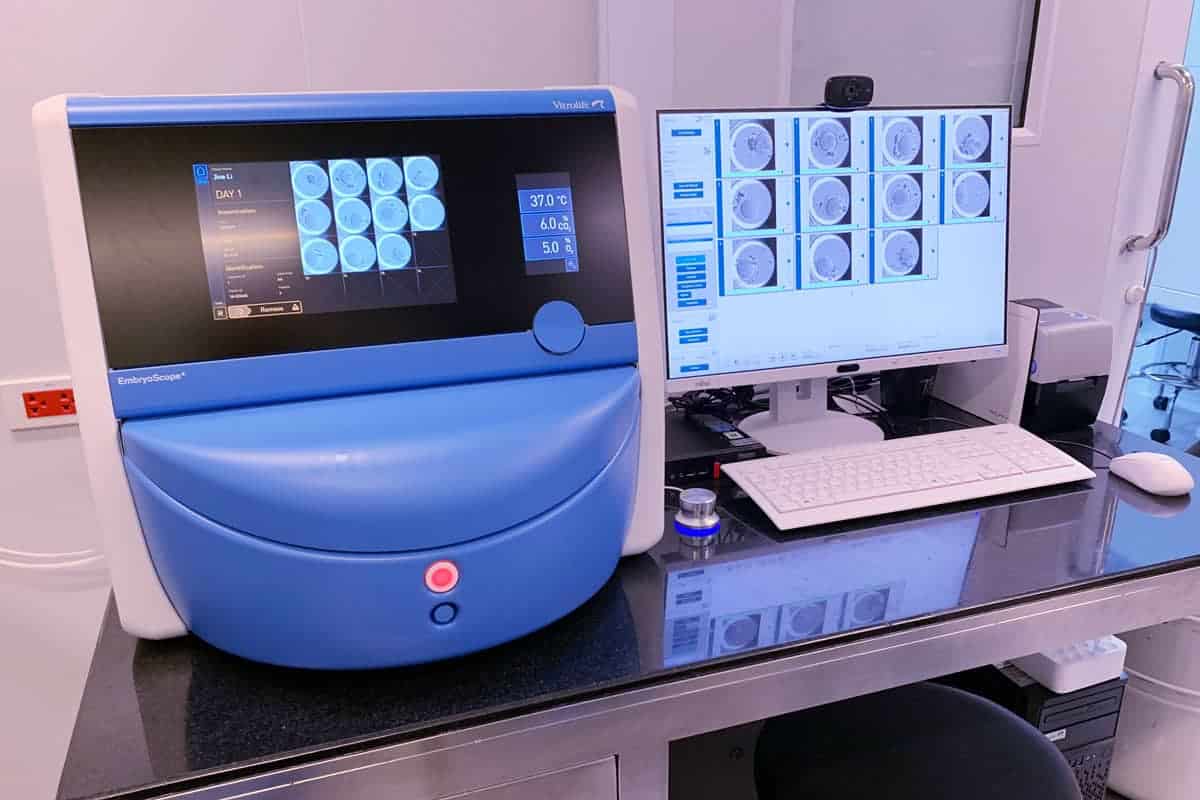
Vejthani Hospital’s VFC Center in Bangkok uses the EmbryoScope Plus System for our in vitro fertilization (IVF) treatments. This state-of-the-art technology monitors embryo development in a time-lapse system that combines an incubator, a high-resolution camera, and computer software.
The system integrates three core components:
- Advanced Embryo Culture Incubator: This incubator fosters a stable and precisely controlled environment, mimicking the ideal conditions for optimal embryo growth.
- Integrated Microscopic Imaging System: This sophisticated system incorporates a high-resolution camera and a specialized stereo microscope. It allows for continuous, noninvasive monitoring of embryo development without the need for manual removal, minimizing potential disruption.
- EmbryoViewer Software: This software program carefully compiles the captured images, generating time-lapse videos that depict the embryo’s development over the culture period.
How The EmbryoScope Plus System Works
Now that you are familiar with the core components, here is how the EmbryoScope Plus system works:
- The embryos are cultured in a special incubator with a built-in microscope and camera.
- The camera takes pictures of the embryos every 10 minutes or so.
- These images are then compiled into a time-lapse video that shows the development of each embryo over time.
- The embryologist can then use this video to assess the health and development of the embryos and select the best ones for transfer.
Benefits of EmbryoScope Plus for IVF
There are several benefits to using the EmbryoScope Plus system:
Undisturbed Culture Environment
The EmbryoScope system eliminates the need to remove the embryos from the incubator for observation, helping to maintain a stable and uninterrupted culture environment, which can improve their chances of development.
Detailed Development Insights And Improved Embryo Selection
The time-lapse technology offered by EmbryoScope Plus provides embryologists with detailed insights into the embryo’s developmental milestones. This continuous monitoring captures crucial moments, such as cell division timings, which were previously difficult to pinpoint. Such detailed observations allow for a more informed selection process, identifying embryos with the highest potential for a successful pregnancy.
Reduced Embryo Stress
Traditional IVF methods require embryos to be removed from the incubator periodically, exposing them to potential environmental stressors like temperature and humidity fluctuations. The EmbryoScope Plus eliminates this need, keeping the embryos in a consistently safe environment, thus enhancing their development and viability.
The Future of IVF with EmbryoScope
As research into time-lapse continues, we can expect further refinements to embryo selection processes and potentially even the identification of new markers for embryo health. This technology holds immense promise for improving IVF outcomes and offering a more personalized and patient-centered experience. Additionally, the system offers patients a less invasive approach and the opportunity to be more involved in their IVF journey.
Visit the VFC Center to Increase Your Chances of Conceiving
At the VFC Center at Vejthani Hospital in Bangkok, we utilize the cutting-edge EmbryoScope Plus technology to significantly increase your chances of a successful IVF outcome. With an impressive success rate of 88.61%, our VFC Center is dedicated to continuously innovating and incorporating new technologies and procedures to assist patients on their path to parenthood.
If you are considering IVF and want to learn more about how the EmbryoScope Plus can be a crucial part of your fertility journey, we invite you to schedule an appointment with our specialists today.